The Road to Recovery: Prognosis and Follow-up in Osteoid Osteoma
Osteoid osteoma, a benign bone tumor, represents a diagnostic and therapeutic challenge due to its varied clinical presentation and potential for significant morbidity. This article provides a comprehensive overview of osteoid osteoma, encompassing its etiology, epidemiology, pathophysiology, clinical manifestations, diagnostic modalities, treatment options, and prognosis.
Etiology and Pathophysiology
Osteoid osteoma typically originates from osteoblasts, the bone-forming cells responsible for bone growth and repair. Histologically, it is characterized by a central nidus composed of vascularized osteoid tissue surrounded by reactive bone formation. The exact etiology of osteoid osteoma remains elusive, although genetic predisposition and local trauma have been implicated as potential contributing factors. Genetic studies have identified mutations in genes related to bone development and regulation, providing insights into the molecular mechanisms underlying tumor formation.
Epidemiology
Osteoid osteoma accounts for approximately 10% of all benign bone tumors and predominantly affects young adults, with a peak incidence in the second and third decades of life. It exhibits a male predominance, with a male-to-female ratio ranging from 2:1 to 5:1. While osteoid osteoma can occur in any bone, it most commonly affects the long bones of the lower extremities, particularly the femur and tibia. However, it can also occur in the spine, with a predilection for the posterior elements.
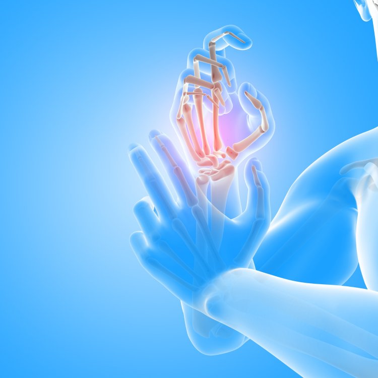
Clinical Manifestations
The clinical presentation of osteoid osteoma varies depending on the size and location of the tumor within the bone. The hallmark symptom is localized pain, which is often described as intense and nocturnal, worsening at night and improving with NSAIDs. Patients may also experience swelling, tenderness, and limited range of motion in the affected area. However, due to its nonspecific symptoms, osteoid osteoma is frequently misdiagnosed initially, leading to delays in appropriate management.
Diagnostic Modalities
Diagnosing osteoid osteoma requires a systematic approach incorporating clinical evaluation, imaging studies, and occasionally, histopathological examination. Conventional radiographs typically demonstrate a radiolucent nidus surrounded by sclerotic bone, although it may be subtle or obscured by overlying structures. Advanced imaging modalities such as CT and MRI offer higher sensitivity and specificity for detecting the nidus, particularly in cases where it is small or located in anatomically complex regions. In cases where the diagnosis is uncertain, a biopsy may be warranted to confirm the presence of osteoid tissue and exclude other differential diagnoses.
Treatment Options
The management of osteoid osteoma depends on several factors, including the location, size, and symptoms associated with the tumor. Conservative management with NSAIDs may provide temporary relief of symptoms; however, it does not address the underlying tumor. Therefore, definitive treatment typically involves surgical excision of the nidus and surrounding reactive bone. Traditional open surgery has historically been employed; however, minimally invasive techniques such as percutaneous radiofrequency ablation (RFA) have gained popularity due to their efficacy, safety, and shorter recovery times. RFA involves the percutaneous insertion of a radiofrequency probe into the nidus under imaging guidance, followed by the application of thermal energy to ablate the tumor while minimizing damage to surrounding healthy tissue.
Prognosis
The prognosis for patients with osteoid osteoma is generally favorable, particularly following successful treatment. Complete excision of the tumor typically results in resolution of symptoms and restoration of normal function. However, recurrence is possible in rare cases, particularly if the nidus is incompletely removed during surgery or if there are multifocal lesions. Long-term follow-up is recommended to monitor for recurrence and ensure optimal outcomes.
Osteoid osteoma poses a diagnostic and therapeutic challenge due to its varied clinical presentation and potential for significant morbidity. However, with advances in imaging technology, minimally invasive techniques, and multidisciplinary collaboration, patients can expect favorable outcomes and restoration of quality of life. Continued research efforts aimed at elucidating the underlying pathophysiology of osteoid osteoma and optimizing treatment strategies are essential to further improve patient outcomes and reduce the burden of this condition.
Disclaimer
The information provided in this article is for educational purposes only and should not be considered medical advice. If you have any health concerns or are experiencing symptoms, it is important to consult with a healthcare professional, such as a doctor or clinic, for proper diagnosis and treatment. Always seek the advice of your doctor or other qualified health provider with any questions you may have regarding a medical condition. Do not disregard professional medical advice or delay in seeking it because of something you have read in this article.
Hashtags
#OsteoidOsteoma #BoneTumors #Orthopedics #Health #MedicalAdvice
What's Your Reaction?