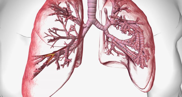
Innovative Therapies for Bronchiectasis: Improving Quality of Life
Bronchiectasis is a chronic pulmonary disorder characterized by irreversible dilation and destruction of the bronchial walls. This condition results from a complex interplay of infectious and non-infectious factors leading to chronic inflammation and airway damage. The consequences of bronchiectasis include impaired mucociliary clearance, recurrent lung infections, and progressive loss of lung function.

Etiology
Bronchiectasis can arise from numerous etiologies, which can be broadly categorized into congenital and acquired causes:
Infectious Causes:
- Bacterial Infections: Recurrent or severe infections such as Mycobacterium tuberculosis, Haemophilus influenzae, Pseudomonas aeruginosa, and Bordetella pertussis can damage the bronchial walls.
- Viral Infections: Severe viral infections, including measles and influenza, can contribute to bronchial damage.
- Fungal Infections: Aspergillosis and other fungal infections may lead to bronchiectasis, particularly in immunocompromised individuals.
Genetic and Congenital Disorders:
- Cystic Fibrosis (CF): The most common genetic cause, CF leads to thick, sticky mucus production that obstructs airways and fosters bacterial growth.
- Primary Ciliary Dyskinesia (PCD): A genetic disorder causing defects in the cilia, resulting in impaired mucus clearance and chronic infections.
- Alpha-1 Antitrypsin Deficiency: This genetic condition can lead to chronic lung disease, including bronchiectasis.
Obstructive Causes:
- Foreign Body Aspiration: Inhaled objects can obstruct airways and cause localized bronchial dilation.
- Tumors: Both benign and malignant tumors can cause bronchial obstruction and subsequent bronchiectasis.
Autoimmune and Inflammatory Diseases:
- Rheumatoid Arthritis: Systemic inflammation can extend to the lungs, causing bronchiectasis.
- Inflammatory Bowel Disease (IBD): Conditions like Crohn's disease and ulcerative colitis are associated with bronchiectasis.
Other Causes:
- Immunodeficiency Disorders: Conditions such as common variable immunodeficiency (CVID) and HIV/AIDS can predispose individuals to recurrent infections and bronchiectasis.
- Environmental and Occupational Exposures: Chronic exposure to pollutants, toxic gases, and dust can contribute to airway damage.
Pathophysiology
The development of bronchiectasis involves a vicious cycle of infection, inflammation, and structural damage. The initial insult, which can be an infection, obstruction, or genetic defect, leads to inflammation and destruction of the bronchial walls. This damage impairs the normal mucociliary clearance mechanism, allowing mucus to accumulate in the airways. The stagnant mucus creates an environment conducive to bacterial colonization and recurrent infections, further exacerbating inflammation and airway damage. Over time, this cycle results in the characteristic dilation and thickening of the bronchi.
Clinical Presentation
Patients with bronchiectasis typically present with a chronic productive cough, often producing large amounts of purulent sputum. Other symptoms may include:
- Dyspnea: Shortness of breath, especially during exertion.
- Recurrent Respiratory Infections: Frequent episodes of bronchitis or pneumonia.
- Hemoptysis: Coughing up blood, which can range from mild to severe.
- Chest Pain: Pleuritic chest pain, which may be exacerbated by coughing.
- Fatigue: Generalized weakness and tiredness.
- Wheezing: Due to airway obstruction and inflammation.
Physical examination findings may include crackles (rales) and wheezing on auscultation, digital clubbing (enlargement of the fingertips), and signs of chronic respiratory distress.
Diagnosis
The diagnosis of bronchiectasis is based on clinical evaluation, imaging studies, and laboratory tests. Key diagnostic tools include:
- High-Resolution Computed Tomography (HRCT) Scan: The gold standard for diagnosing bronchiectasis. HRCT can reveal bronchial dilation, wall thickening, mucus plugging, and other structural abnormalities.
- Pulmonary Function Tests (PFTs): These tests assess the degree of airflow obstruction and restriction, often showing a pattern of obstructive lung disease.
- Sputum Culture and Sensitivity: Identifying pathogens in the sputum helps guide antibiotic therapy. Chronic bacterial colonization, such as with Pseudomonas aeruginosa, is common.
- Blood Tests: Complete blood count (CBC) and tests for underlying conditions, such as immunodeficiency or autoimmune diseases, can be useful.
- Bronchoscopy: This procedure may be used to evaluate airway abnormalities, obtain samples for culture, and remove obstructions.
Treatment
Management of bronchiectasis focuses on controlling infections, reducing inflammation, and improving mucus clearance. Treatment options include:
- Antibiotics: To treat acute exacerbations and prevent infections. Long-term antibiotics, such as azithromycin, may be used in cases of frequent exacerbations.
- Bronchodilators: To relieve airway obstruction and improve airflow. These include beta-agonists and anticholinergic agents.
- Mucolytics and Hypertonic Saline: To help thin and mobilize mucus, making it easier to clear from the airways.
- Chest Physiotherapy: Techniques such as postural drainage, percussion, and vibration to aid mucus clearance.
- Inhaled Corticosteroids: To reduce airway inflammation in some patients.
- Surgical Intervention: In severe or localized cases, surgical resection of the affected lung segments or lobes may be considered.
- Vaccinations: Annual influenza and pneumococcal vaccines to reduce the risk of respiratory infections.
Prognosis
The prognosis for patients with bronchiectasis varies depending on the underlying cause, severity of the disease, and the effectiveness of treatment. With proper management, many patients can lead a relatively normal life. However, severe cases may lead to significant morbidity, frequent hospitalizations, and decreased quality of life. Complications of bronchiectasis include respiratory failure, cor pulmonale (right-sided heart failure), and, rarely, massive hemoptysis.
Bronchiectasis is a chronic and potentially debilitating respiratory condition that requires careful management to prevent complications and improve patient outcomes. Early diagnosis and a comprehensive treatment approach are essential in managing the disease and enhancing the quality of life for affected individuals. Ongoing research and advances in medical treatments hold promise for better management and outcomes for patients with bronchiectasis.
Disclaimer: The information provided in this article is for educational purposes only and should not be considered medical advice. If you have any health concerns or are experiencing symptoms, it is important to consult with a healthcare professional, such as a doctor or clinic, for proper diagnosis and treatment. Always seek the advice of your doctor or other qualified health provider with any questions you may have regarding a medical condition. Do not disregard professional medical advice or delay in seeking it because of something you have read in this article.
#hashtags: #Bronchiectasis #RespiratoryHealth #ChronicIllness #MedicalEducation #Healthcare
What's Your Reaction?


