Drooping Eyelids: Causes, Symptoms, and Cutting-Edge Treatments for Ptosis
Ptosis, also known as blepharoptosis, is the medical term for drooping of the upper eyelid. This condition can affect one or both eyes and can be present at birth (congenital) or develop later in life (acquired). Ptosis can range from a slight droop to a complete closure of the eyelid, which can obstruct vision and affect daily activities. Understanding the causes, symptoms, and treatment options for ptosis is crucial for effective management.
Causes of Ptosis
Congenital Ptosis:
- Developmental Issues: Congenital ptosis is often due to the underdevelopment or dysfunction of the levator palpebrae superioris muscle, responsible for lifting the eyelid.
- Genetic Factors: It may be inherited and linked to genetic disorders such as congenital myopathies or neurofibromatosis.
- Associated Syndromes: Conditions like Marcus Gunn jaw-winking syndrome, where eyelid movement is linked to jaw movement, can be associated with congenital ptosis.
Acquired Ptosis:
- Neurogenic Ptosis:
- Third Nerve Palsy: Damage to the oculomotor nerve (cranial nerve III) can cause ptosis, often accompanied by eye movement abnormalities and pupil dilation.
- Horner's Syndrome: Disruption of the sympathetic nerve pathway can lead to mild ptosis, pupil constriction (miosis), and anhidrosis (lack of sweating) on the affected side.
- Myogenic Ptosis:
- Myasthenia Gravis: An autoimmune disease causing weakness in the voluntary muscles, including the eyelid muscles, leading to fluctuating ptosis.
- Muscular Dystrophies: Genetic disorders that weaken muscle fibers, such as oculopharyngeal muscular dystrophy.
- Aponeurotic Ptosis:
- Age-Related Changes: The levator aponeurosis, a fibrous tissue that connects the levator muscle to the eyelid, can weaken or stretch with age.
- Post-Surgical: After eye surgeries like cataract removal, the levator aponeurosis can be inadvertently affected, leading to ptosis.
- Mechanical Ptosis:
- Eyelid Tumors: Benign or malignant growths can weigh down the eyelid.
- Severe Edema: Swelling due to infection or inflammation can cause the eyelid to droop.
- Traumatic Ptosis: Injury to the eyelid or surrounding structures can damage the muscles or nerves responsible for eyelid elevation.
Symptoms of Ptosis
- Drooping Eyelid: The most apparent symptom, which can vary in severity.
- Obstructed Vision: If the drooping eyelid covers part or all of the pupil.
- Eye Strain and Fatigue: Resulting from efforts to keep the eyes open or compensate for the drooping lid.
- Forehead Wrinkling: Due to the use of forehead muscles to lift the eyelids.
- Chin-Up Posture: Tilting the head back to see under the drooping eyelid.
- Double Vision: In cases where ptosis is associated with other ocular abnormalities.
- Compensatory Mechanisms: Such as raising the eyebrows to lift the eyelid.
- In children with congenital ptosis, additional symptoms may include amblyopia (lazy eye), astigmatism, or developmental delays in vision.
Diagnosis of Ptosis
A comprehensive eye examination by an ophthalmologist is essential for diagnosing ptosis. The evaluation typically includes:
- Medical History: Detailed review of the patient’s medical and family history.
- Physical Examination: Observing the eyelid position, symmetry, and movement.
- Eyelid Measurements: Assessing the degree of drooping, levator function, and margin-reflex distance (distance between the upper eyelid margin and the light reflex on the cornea).
- Visual Acuity Tests: Checking for vision impairment caused by ptosis.
- Pupil Examination: To rule out associated neurological conditions.
- Fatigue Testing: For conditions like myasthenia gravis, where ptosis worsens with prolonged use of the eyelid muscles.
- Imaging Studies: MRI or CT scans if a tumor or neurological cause is suspected.
- Electromyography (EMG): To assess muscle function if a myogenic cause is suspected.
- Blood Tests: To identify underlying conditions like myasthenia gravis.
Treatment of Ptosis
Treatment depends on the severity and underlying cause of ptosis. Options include:
- Observation: In mild cases where vision is not significantly affected, regular monitoring may be adequate.
- Nonsurgical Treatments:
- Medications: For ptosis caused by myasthenia gravis, anticholinesterase agents or immunosuppressants can be effective.
- Botulinum Toxin (Botox): In some cases, Botox injections can help improve muscle function temporarily.
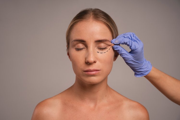
- Surgical Treatments:
- Levator Resection or Advancement: Involves tightening or shortening the levator muscle to elevate the eyelid. This is the most common surgical approach.
- Frontalis Sling Procedure: Utilizes a sling material (often silicone or fascia) to connect the eyelid to the frontalis muscle (forehead muscle), allowing the forehead muscle to lift the eyelid. This is often used in severe ptosis or when the levator muscle is non-functional.
- Müller's Muscle-Conjunctival Resection: A procedure where a portion of Müller's muscle and conjunctiva is removed to elevate the eyelid, suitable for mild to moderate ptosis with good levator function.
- External Levator Aponeurosis Advancement: Involves advancing the levator aponeurosis to its normal position to improve eyelid height.
- Non-Surgical Devices:
- Ptosis Crutches: Small devices attached to glasses that mechanically lift the eyelid. These are suitable for patients who cannot undergo surgery.
Postoperative Care and Complications
Post-surgery, patients may experience bruising, swelling, and temporary discomfort. Proper postoperative care includes:
- Follow-Up Visits: Regular check-ups with the ophthalmologist to monitor healing and ensure the desired outcome.
- Medications: Prescribed antibiotics or anti-inflammatory medications to prevent infection and reduce swelling.
- Activity Restrictions: Avoiding strenuous activities and following specific guidelines for eye care to promote healing.
Potential complications from ptosis surgery include:
- Under-Correction or Over-Correction: The eyelid may not be lifted enough or may be lifted too much.
- Asymmetry: Differences in eyelid height between the two eyes.
- Dry Eyes or Excessive Tearing: Due to altered eyelid position affecting tear distribution.
- Infection or Scarring: As with any surgical procedure, there is a risk of infection and scarring.
Disclaimer: The information provided in this article is for educational purposes only and should not be considered medical advice. If you have any health concerns or are experiencing symptoms, it is important to consult with a healthcare professional, such as a doctor or clinic, for proper diagnosis and treatment. Always seek the advice of your doctor or other qualified health provider with any questions you may have regarding a medical condition. Do not disregard professional medical advice or delay in seeking it because of something you have read in this article.
Hashtags: #Ptosis #EyeHealth #MedicalEducation #EyelidSurgery
What's Your Reaction?